The lingual foramen is a small midline opening on the posterior aspect of the symphysis of the mandible just above the mental spine. PDF On Jan 1 2020 K.

Normal Radiographic Anatomical Landmarks
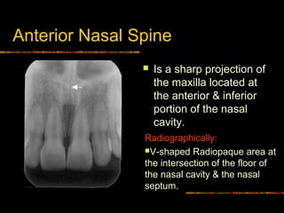
Radiopaque anatomical landmarks - Nasal septum Floor of nasal cavity Anterior nasal spine Inferior nasal conchae.

. It represents the anterior continuation of the mandibular canal. Incisive foramen is the opening of the incisive canal located immediately behind the maxillary central incisors. The mandibular incisive canal MIC is a small bony channel located in the interforaminal region.
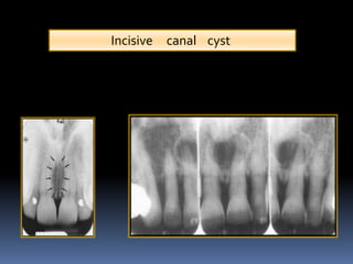
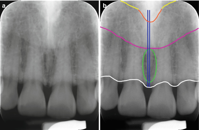
Incisive foramen RadiographPhotograph Frame 22. Purple ellipseinferior nasal concha. An incisive canal cyst is a developmental cyst non neoplastic cyst arising from degeneration of nasopalatine ducts.
Nerve and artery enter posterior alveolar foramen on the infratemporal surface of the maxilla Passes in the maxilla anteriorly giving off branches to the teeth bone and gingiva what are nutrient canals and what do they look like. Incisive Fossa Radiograph - 12 images - canine radiographs normal oral radiographic anatomy head and neck bones anatomy on radiographs intraoral radiographs part i. The persistence of ductal epithelium leads to formation of cyst.
Light green ellipselarge incisive foramen. The incisive nerve innervates the anterior palatal soft tissues. A b Maxillary occlusal radiograph.
Here a posterior-occlusal view of a skull demonstrates the incisive foramen. Yellowfloor of the nasal cavity. The mean width of the foramen labiopalatally and mesiodistally was 312 094 mm and 323 098 mm respectively.
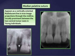
Maxilla Incisor region Radiolucent anatomical landmarks - Incisive foramen Median palatal suture Nasal cavity Superior foramina of the Nasopalatine canal Lateral fossa. Foramen maxillary mand radiology interpretation max maxilla central arrows. Maxillary premolar region The radiographs which were taken from premolar region exhibit the floor of the nasal cavity and maxillary sinus usually separated from septum above the root tip of the second premolar Fig3C.
The incisive foramen although. It is considered the most common non-odontogenic cyst and develops only in the midline anterior maxilla. Inverted Y formation Radiograph comparison Frame 27.
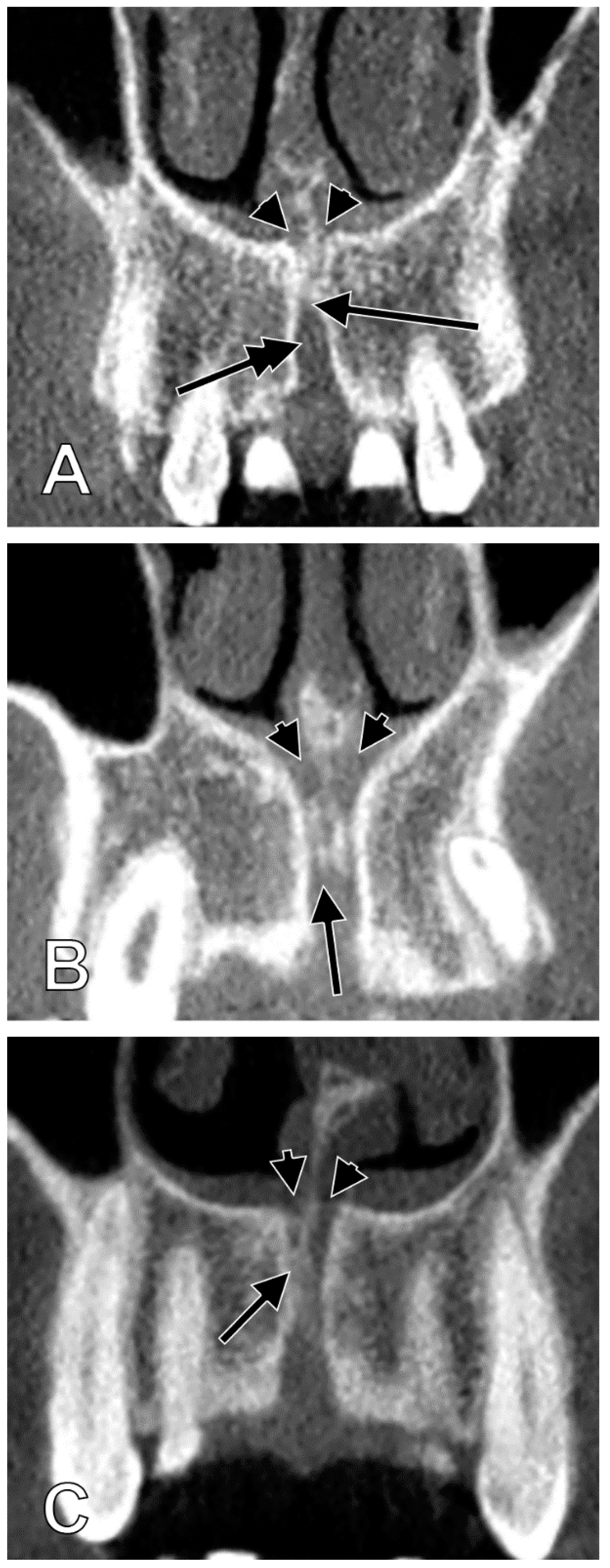
The incisive foramen also known as nasopalatine foramen or anterior palatine foramen is the oral opening of the nasopalatine canal. Anterior extent of maxillary sinus Radiograph Frame 25. On periapical x-ray images the incisive foramen is located in the midline between the roots of the central incisors.
These ducts usually regress in fetal life. Maxillary sinus Illustration Frame 23. Incisive foramen Nasal septum Radiograph Frame 21.
It is located in the maxilla in the incisive fossa midline in the palate posterior to the central incisors at the junction of the medial palatine and incisive sutures. Sometimes these bone canals mimic a dentino- or osteolysis on conventional. The total percentage of visibility of the incisive foramen in the bisecting and paralleling radiographs was 40 and 767 respectivelyIt is important to take into account the relationship between the structures the aortic root encompasses.
Veena and others published Appreciation of Incisive Foramen in Intraoral Periapical Radiographs - A Comparative Radiographic Study. Maxillary sinus Radiograph Frame 24. It can be single or multiple.
Article on Appreciation of Incisive Foramen in Intraoral Periapical Radiographs - A Comparative Radiographic Study published in Journal of Morphological Sciences 38 on 2021-01-01 by K. Paired structures are marked on one side only. Read the article Appreciation of Incisive Foramen in Intraoral Periapical Radiographs - A Comparative Radiographic Study on R Discovery your go-to avenue for.
There is widespread knowledge that the mental foramen or incisive foramen can be projected onto tooth roots in conventional radiographs thus simulating apical lesions. White ellipseair in nasal cavity. The incisive foramen generally appears in most panoramic radiographs though not with the clarity seen in periapical radiographs.
Mean canal length was 1863 235 mm and males. Changes in the size of one structure cause the improper functioning of the adjacent ones. Lingual foramen - Wikipedia Lingual foramen The small lingual foramen black hole in lower portion of picture as seen on a periapical radiograph of the anterior mandible.
Its appearance is quite variable due to normal anatomic variation and due to the operators angulation of the x-ray beam. Nasopalatine canal incisive foramen and anterior lobe of the maxillary sinus can also be visible in this projection Fig3B. Inverted Y formation Radiograph Frame 26.
The pear-shaped radiolucency between the apices of the central incisors. In contrast the anatomical structure of the canalis sinuosus and its branching canals in the anterior maxilla are less known. Assessments included 1 mesiodistal diameter 2 labiopalatal diameter 3 length of the incisive canal 4 shape of incisive canal and 5 width of the bone anterior to the incisive foramen.
Dark bluemidline palatal suture. Light bluewall of maxillary sinus.

Mouth Incisive Canal Cyst Professional Radiology Outcomes

Visibility Of Mandibular Anatomical Landmarks In Panoramic Radiography A Retrospective Study Semantic Scholar

Pdf The Evaluation Of Visibility Of Mandibular Anatomic Landmarks Using Panoramic Radiography Semantic Scholar
Normal Radiographic Anatomical Landmarks
Normal Anatomical Landmarks In Dental X Rays And Cbct Springerlink

Panoramic Radiograph Showing Mandibular Incisive Canal Arrow Download Scientific Diagram

The Appearance Of The Mental Foramen On Panoramic Radiographs Download Scientific Diagram

Measurement Of Incisive Foramen Blue Line Nasal Foramen Red Line Download Scientific Diagram

Periapical Radiograph 1 Year After Treatment Bone And Teeth Showing Download Scientific Diagram

6 Essentials Of Dental Radiographic Analysis And Interpretation Pocket Dentistry
Intra Oral Radiographic Anatomical Landmarks

Incisive Foramen Dr G S Toothpix

Pdf Visibility Of Maxillary And Mandibular Anatomical Landmarks In Digital Panoramic Radiographs A Retrospective Study Semantic Scholar
Normal Radiographic Anatomical Landmarks

Figure 2 Assessment Of The Mandibular Incisive Canal By Panoramic Radiograph And Cone Beam Computed Tomography

Opg Showing Incisive Foramen And Mental Foramen Download Scientific Diagram

Radiographic Interpretation In Oral Medicine And Hospital Dental Practice Pocket Dentistry
Anatomia Free Full Text Detailed Morphology Of The Incisive Or Nasopalatine Canal Html

